| |
p53 database
p53 database
Database curation
Download the database
Improve p53 mutation detection and report
p53 mutation and cancer
|
p53 MUTATIONS IN BRAIN CANCER
Brain tumors
Tumors that begin in brain tissue are known as primary brain tumors. Primary brain tumors are classified by the type of tissue in which they begin. The most common brain tumors are gliomas, which begin in the glial (supportive) tissue. There are several types of gliomas:
* Astrocytomas arise from small, star-shaped cells called astrocytes. They may grow anywhere in the brain or spinal cord. In adults, astrocytomas most often arise in the cerebrum. In children, they occur in the brain stem, the cerebrum, and the cerebellum. A grade III astrocytoma is sometimes called anaplastic astrocytoma. A grade IV astrocytoma is usually called glioblastoma multiforme.
* Brain stem gliomas occur in the lowest, stemlike part of the brain. The brain stem controls many vital functions. Tumors in this area generally cannot be removed. Most brain stem gliomas are high-grade astrocytomas.
* Ependymomas usually develop in the lining of the ventricles. They may also occur in the spinal cord. Although these tumors can develop at any age, they are most common in childhood and adolescence.
* Oligodendrogliomas arise in the cells that produce myelin, the fatty covering that protects nerves. These tumors usually arise in the cerebrum. They grow slowly and usually do not spread into surrounding brain tissue. Oligodendrogliomas are rare. They occur most often in middle-aged adults but have been found in people of all ages.
More information can be found at: http://neurosurgery.mgh.harvard.edu/newwhobt.htm
Spectrum of p53 mutations in brain cancer
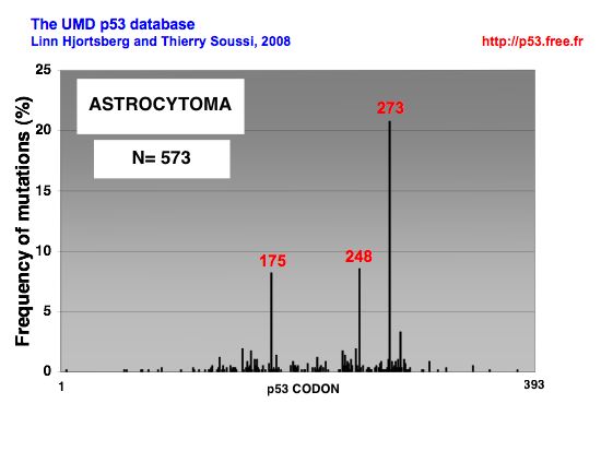 |
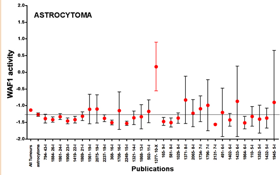
DISTRIBUTION OF p53 MUTATIONS IN ASTROCYTOMA |
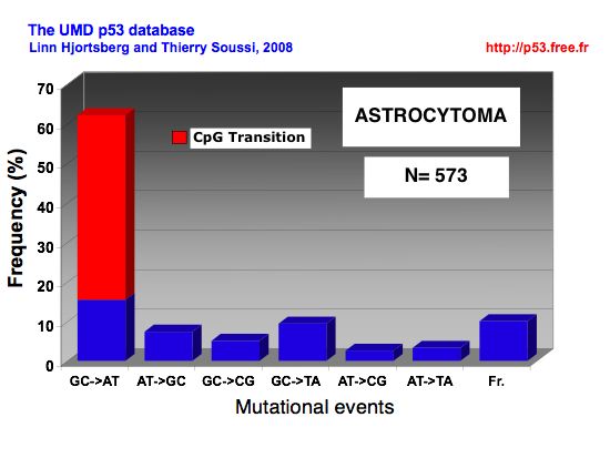 |
MUTATIONAL EVENTS IN ASTROCYTOMA |
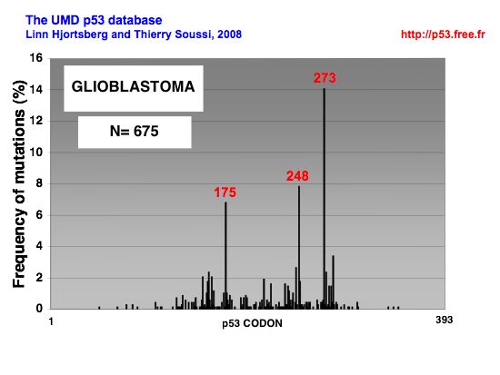 |
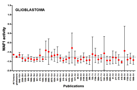
DISTRIBUTION OF p53 MUTATIONS IN GLIOBLASTOMA |
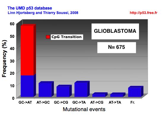 |
MUTATIONAL EVENTS IN GLIOBLASTOMA |
|
|
Meta-analysis of p53 loss of function in bladder cancer.
Points ; mean p53 activity as measured by transactivation with the WAF1promoter ; bars , 95 % CI. The mean and 95 % CI of p53 activity for all studies combined for a specific type of cancer is shown on the far left of each graph.
Horizontal line, mean of the combined studies. The publication code is indicated on the x-axis : the first number is an anonymous ID for the publication and the second number indicates the number of p53 mutants included in this study. Studies are presented from left to right in decreasing order of number of p53 mutants. The y-axis corresponds to p53 transactivation activity, with a value of 1.5 for the negative control and a value of 2.5 for 100 % of wild-type activity. Only studies with 5 or more p53 mutations are shown on the graph.
More information about this statistical analysis can be found in this article:
Soussi, T., Asselain, B., Hamroun, D., Kato, S., Ishioka, C., Claustres, M. and Beroud, C. (2006) Meta-analysis of the p53 mutation database for mutant p53 biological activity reveals a methodologic bias in mutation detection. Clin Cancer Res, 12, 62-69. Download the pdf |
|
|