Our Work

Our work

p53 structure

Xenopus p53

p53 in Lung cancer

p53 in Breast cancer

p53 antibodies in cancer

p73 antibodies in cancer

Publications of the lab

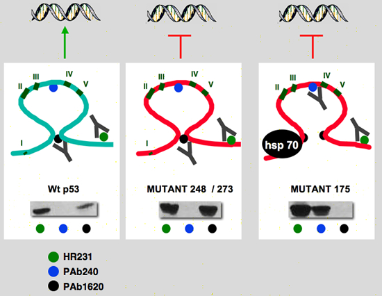
Structure – function studies of the p53 protein

The main objective of our work is to understand the role of mutant p53 in human tumorigenesis. Mutations in the gene of the tumor suppressor p53 represent the most frequent genetic alterations in human cancer, affecting about 50% of all individual tumors. The major effect of these mutations is the elimination of the various tumor suppressing functions demonstrated for wild-type p53. In addition, there is accumulating evidence for an active role of mutant p53 in tumorigenesis, resulting from a "gain of function" for at least some mutant p53 proteins. Mutant p53 gain of function becomes evident because of an increased tumorigenic potential of tumor cells expressing mutant p53 genes, as well as alterations in growth properties of such cells in culture. Furthermore, data available indicate that the presence of certain p53 mutations in several types of human cancer correlates with less favourable patient prognosis. Based on these observations, we have undertaken the analysis of the heterogeneityof mutant p53.

Some mutant p53 display a change of conformation

The structural difference between the various mutant TP53 was initially identified using monoclonal antibodies able to discriminate mutations that change TP53 folding and mutations in the residues involved in DNA recognition (Gannon et al., 1990; Legros et al., 1994). Two classes of mutations have been distinguished on the basis of various in vitro assays and the three-dimensional structure of the protein (Cho et al., 1994):
class I mutations, exemplified by mutants at codon 248 (7.6% in the p53 database,http://p53.free.fr/, 2005 release), affect amino acids directly involved in the protein-DNA interaction. They have a wild-type conformation as probed by conformational monoclonal antibodies and they do not bind to the chaperone hsp70 (Hinds et al., 1990; Ory et al., 1994).
Class II mutations, exemplified by the mutant at codon 175 (4.9% in the database), have an altered conformation with intense binding to hsp70. The amino acids altered in this class of mutants are involved in stabilizing the tertiary structure of the protein. Class II mutations are associated with a more severe phenotype in vitro than class I mutations (Ory et al., 1994).

p53_structure
Conformational model of p53. Wild-type p53 has a very compact structure which is recognized by Pab1620 but not by HP64 or HO3.5. The presence of a mutation induces relaxation of this structure which inactivates the epitope for PAB1620 but renders the epitopes for HP64 and HO3.5 accessible (from Legros et al., 1994; Ory et al., 1994).

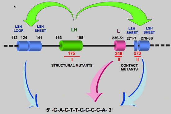
The determination of the 3-dimensional crystal structure of p53 has also shed light on these two classes of mutants. As shown in Figure 6, crystallography of the central region of p53 with its DNA recognition motif has allowed a very precise definition of this interaction. Two types of regions have been identified :
i) regions containing amino acids in direct contact with DNA (regions 112-141, 236-251 and 271-286) and ii) a region critical for maintenance of the tertiary architecture of the central domain of p53 (region 163-195) which joins the DNA binding regions.

p53_structure

Class II mutants correspond to the residues in direct contact with DNA (contact mutants) and do not cause large alterations of the spatial structure of p53.

Class I mutants affect the amino acids required for maintenance of p53 structure (conformational mutants) and completely destructure the p53 conformation. This is why the cryptic epitope recognized by the monoclonal antibody HP64 is detected in the mutant proteins. Furthermore, this allows better understanding of the interaction of some mutant p53s with HSP70, which recognize hydrophobic motifs inaccessible in the native form. These two types of regions corresponded to the two class of mutants we had previously defined :

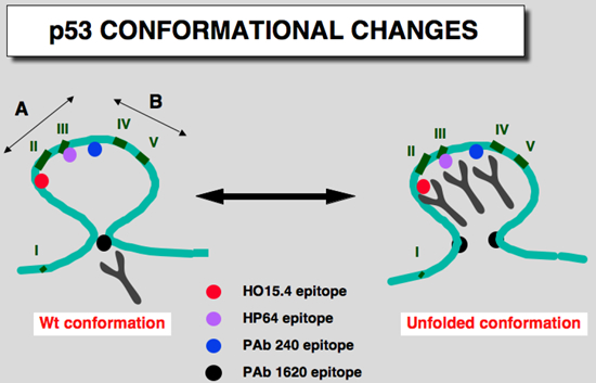
Conformational studies with a new set of p53 monoclonal antibodies

Using a set of overlapping peptides of the human p53 protein, we analysed the epitopes recognized by 18 monoclonal antibodies specific for human p53 , that were produced in our laboratory. We showed that most of these epitopes corresponded to linear antigenic determinants which lie predominantly in the amino- or carboxy-terminus of the p53 protein.

Using a truncated p53 (residues 66 to 361), we selected eight new monoclonal antibodies directed to thecentral part of the protein. We then identified the epitopes recognized by seven out of these eight antibodies with a set of overlapping peptides. One of these antibodies had an epitope similar to PAb240, whereas the others recognized novel and diverse antigenic determinants. Using a series of 19 p53 mutants, we showed that the behavior of several of the new monoclonal antibodies was similar to that of PAb240 despite their various epitope localizations.
This suggests that different mutations in the p53 protein induce an overall conformational change that can be detected by various monoclonal antibodies directed toward the central part of the protein.

p53_structure
A model for p53 confomation: The amino and carboxy-termini of th p53 protein are highly acessible and are displayed at the surfaceof the prolein. They contain the immunodominant regjons. The central region is ralher compact and allows the formation of he epitope for PAb1620, but epitopes of PAb240, HO15.4, HO3.5, HO13.1 and HP64 are masked. The presence of a specific mutation or the partial denaturation of the p53 protein destroyed the PAb1620 epitope, unmasked those
localized in the central region of the protein.
I to V corresponds ro the highly conserved domains of the p53 protein (Soussi et al, 1990). This model is based on the conformational nodel proposd by Milner (1991)


Use of hybrids proteins to solve the function of the DNA binding domain in the gain of function of mutant p53 and how it is involved in the conformational change of p53

check the xenopus page


Home | Our Work |p53 Info| p53 Database | p53 Link | Contact us