Our WorkOur workp53 structureXenopus p53p53 in Lung cancerp53 in Breast cancerp53 antibodies in cancerp73 antibodies in cancerPublications of the lab
|
p53 Antibodies in cancer patientsThe latest review on p53 antibodies with the compilation of every studies published from 1989 to 1999. INTRODUCTIONA fundamental characteristic of malignant cells is the accumulation of genetic alterations during tumor progression. Altered genes may not only lead to a functional change that contributes to the appearance of a malignant phenotype but may also generate molecules that will induce humoral or cell mediated specific immune responses. Tumors cells can also express differentiation antigens usually present on certain normal embryonic cells such as a-fetoprotein which is produced by liver cancer cells, and carcinoembryonic antigen (CEA), produced by colon cancer cells and other epithelial tumors. Mutated molecules as well as abnormal expression of differentiation antigens can lead to the development of specific CTL and/or to the appearance of circulating antibodies. In recent years, increasing evidence has accumulated that p53 protein triggers such an immune response. Mutation of the p53 tumor suppressor gene is the most frequent abnormality in various human tumors. More than 95% of these alterations are missense mutations which are scattered in the central part of the gene. Although all these mutations lead to the inactivation of the biological properties of the p53 protein, they also have dramatic consequences in term of p53 stability. Mutant p53 protein, which takes on an abnormal conformation, is more stable than the wild-type (half-life of several hours compared to 20 minutes for the wild type p53), accumulates in the nucleus of neoplastic cells and thus becomes immunologically detectable. An important consequence of this phenomenon is that positive immunostaining is indicative of abnormalities of the p53 gene and its product (Dowell et al., 1994). The recent discovery that p53 mutations can lead to the accumulation of the p53 gene product in tumor cells has shed new light on the earlier observation that anti-p53 antibodies (p53-Ab) are present in sera of patients with breast cancer. While raising the question of the relationship between p53 gene mutation, p53 accumulation and the anti-p53 humoral response, it also opens the way to the development of new cancer diagnostic tools, as discussed below. HISTORICAL NOTESIn 1979, DeLeo et al. showed that the humoral response of mice to some methylcholanthrene-induced tumor cells such as MethA was directed to the p53 protein (De Leo et al., 1979). It was later found that animals bearing several types of tumors elicited an immune response specific for p53 (Kress et al., 1979; Melero et al., 1979; Rotter et al., 1980). In 1982, Crawford et al. first described antibodies against human p53 protein in 9% of breast cancer patient sera (Crawford et al., 1982). No significant clinical correlation was reported, and at that time, no information was available concerning mutations of the p53 gene. Caron de Fromentel et al. later found that such antibodies were present in sera of children with a wide variety of cancers. The average frequency was 12%, but this figure increased to 20 % in Burkitt lymphoma (Caron de Fromentel et al., 1987). Those studies, performed in the early eighties, were virtually ignored for more than 10 years due to the lack of interest in p53 during that period. In the early nineties, it was discovered that the p53 gene is the most common target for molecular alteration in every type of human cancer. This provoked considerable interest in the study of the p53 protein and its function in normal and transformed cells and to the rediscovery of this humoral response which had been found in cancer patients. All the studies described below concern p53-Abs detected in patients at the time of diagnosis, prior to any treatment such as surgery or chemotherapy. p53 ANTIBODY ANALYSISUsing either immunoprecipitation or Western blot, earlier works focused on small series and demonstrated that p53-Ab were detected in patients with various types of cancer (Crawford et al., 1982; Caron de Fromentel et al., 1987). More recently, several ELISA have been developed, enabling the evaluation of large series of patients (Angelopoulou et al., 1994; Lubin et al., 1995). In a series of more than 1000 sera, it was shown that p53-Ab are very rare in healthy donors (less than 0.5%), whereas the frequency of p53-Ab in patients with neoplasias is strictly correlated with the frequency of p53 mutations (Lubin et al., 1995). Most patients with p53-Ab exhibit an accumulation of mutant p53 in their tumor cells (Winter et al., 1992; Wild et al., 1995). However, some patients have p53-Ab, yet no p53 mutation can be detected in the tumor. Whether p53 mutations do exist but for some reason are not detected remains to be established. At this point, it should be emphasized that assay of p53-Ab corresponds to a global approach to assessing p53 alterations and does not depend on tumor sampling, which may be very heterogeneous. In contrast, molecular analysis of tumor tissues or biopsies corresponds to local analysis of p53 status and may be erroneous when the tumor is heterogeneous or highly contaminated by cells from normal tissue.
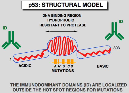
It is also clear that not all patients with a p53 alteration develop p53-Ab. Davidoff et al. suggested that the type of mutation could influence the production of antibodies (Davidoff et al., 1992) but in light of more recent results showing that similar mutations can have a differing behaviors [Preudhomme et al., 1994; Wild etal., 1995; Guinee et al., 1995; Winter et al., 1992), this hypothesis will require further investigation. It is also possible that, for an identical mutation, the immune response is dependent on the specific combination of major histocompatibility complex (MHC) class I and II molecules expressed by each individual. Comparison of the frequency of p53 alterations in the literature indicates that 30 to 40 % of patients with an alteration in the p53 gene develop p53-Ab (Lubin et al., 1995). p53 ANTIBODIES RECOGNIZE LINEAR IMMUNODOMINANT EPITOPESUsing either truncated p53 or synthetic peptides, it has been shown that p53-Ab strongly binds the amino- and carboxy-terminus of the p53 protein, a region which is totally devoid of mutations (Schlichtholz et al., 1992; Lubin et al., 1993). This finding is in agreement with the observation that these p53-Ab recognized wild type or mutants p53 in a similar way (Schlichtholz et al., 1992; Labrecque et al., 1993).. This epitope localization was found to be similar in animals hyperimmunized with human p53 (Legros et al., 1994). Analysis of monoclonal antibodies produced against human p53 demonstrate a strong bias in the epitope recognized by these monoclonal antibodies. Most of them recognized epitopes localized in the amino-terminus of p53. It has been shown that murine or rabbit sera hyperimmunized with human p53 predominantly recognized the amino- and carboxy-terminus of p53 (Legros et al., 1994). Taken together, (i) the correlation between p53 accumulation (and p53 gene mutation) in tumor cells and the p53 antibody response, (ii) the presence of immunodominant epitopes outside the hot spot region of the p53 mutation, (iii) the similarity of the humoral response in patients independent of the cancer type, and (iv) the similarity of the antigenic site profiles between patients and hyperimmunized animals, all suggest that p53 accumulation is a major trigger for the development of a humoral response in cancer patients. As stated above, the level of p53 proteins in a normal organism is very low, suggesting very weak (if any) tolerance to endogenous p53. The accumulation of p53 in tumors could lead to a self-immunization process culminating in the appearance of p53-Ab. Isotyping of these p53-Ab has shown that they correspond mainly to IgG1 and IgG2 subclasses, while some patients exhibit a predominant IgA response (Lubin et al., 1995). Some patients also had IgM, although none had IgM p53-Ab as the only isotype. No IgG3 or IgG4 were detected in this study. Again, this result strengthens the hypothesis of an active humoral response to p53.
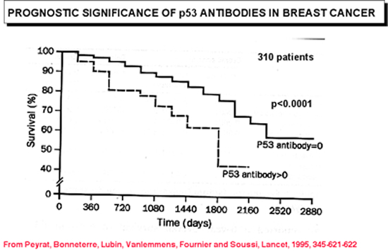
ANTI p53 ANTIBODIES IN BREAST CANCERIn a first step, we have demonstrated that the presence of these antibodies in breast cancer patients is correlated with other indicators of a poor prognosis such as absence of estrogen receptors or histoprognostic grade (Scarff-Bloom-Richardson). An identical correlation has also been described by other authors but by using either a molecular or immunohistochemical approach. More recently we have shown that overall survival in seropositive patients was significantly lower than in seronegative patients.
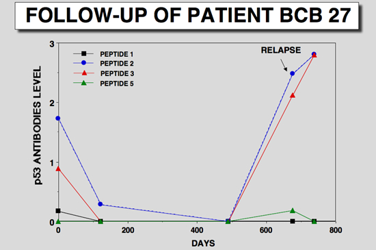
Anti-p53 antibodies can also be used to monitor patients during treatment. Figure below shows an example of a breast cancer patient in which the reappearance of anti-p53 antibodies precedes the detection of relapse.
p53 ANTIBODIES AND LUNG CANCERRecently, p53 antibodies were detected in sera of two patients who were heavy smoker without diagnosed lung malignancy. Both of these patients developed invasive squamous lung cancer, respectively, 5 and 15 months after detection of serum p53 antibodies. In the second patient, p53 overexpression was detected in tumoral cells from bronchial biopsy specimens
Since p53 alterations represent the most common, earliest genetic changes in lung carcinogenesis, it is suggested that p53-Ab detection represents a new and sensitive tool for detection of preneoplastic and microinvasive bronchial lesions in patients with a high risk of lung cancer, i.e., heavy smokers. REFERENCES• De Leo, A. B., Jay, G., Appella, E., Dubois, G. C., Law, L. W., and Old, L. J. (1979). Detection of a transformation-related antigen in chemically induced sarcomas and other transformed cells of the mouse. Proc Natl Acad Sci USA 76, 2420-2424. • Kress, M., May, E., Cassingena, R., and May, P. (1979). Simian Virus 40-transformed cells express new species of proteins precipitable by anti-simian virus 40 serum. J. Virol. 31, 472-483. • Melero, J. A., Stitt, D. T., Mangel, W. F., and Carroll, R. B. (1979). Identification of new polypeptide species (48-55K) immunoprecipitable by antiserum to purified large T antigen and present in simian virus 40-infected and transformed cells. J. Virol. 93, 466-480. • Rotter, V., Witte, O. N., Coffman, R., and Baltimore, D. (1980). Abelson murine leukemia virus-induced tumors elicit antibodies against a host cell protein, p50. J Virol 36, 547-555. • Crawford, L. V., Pim, D. C., and Bulbrook, R. D. (1982). Detection of antibodies against the cellular protein p53 in sera from patients with breast cancer. Int. J. Cancer 30, 403-408. • Caron de Fromentel, C., May-Levin, F., Mouriesse, H., Lemerle, J., Chandrasekaran, K., and May, P. (1987). Presence of circulating antibodies against cellular protein p53 in a notable proportion of children with B-cell lymphoma. Int. J. Cancer 39, 185-189. • Davidoff, A. M., Iglehart, J. D., and Marks, J. R. (1992). Immune response to p53 is dependent upon p53/HSP70 complexes in breast cancers. Proc Natl Acad Sci USA 89, 3439-3442. • Schlichtholz, B., Legros, Y., Gillet, D., Gaillard, C., Marty, M., Lane, D., Calvo, F., and Soussi, T. (1992). The immune response to p53 in breast cancer patients is directed against immunodominant epitopes unrelated to the mutational hot spot. Cancer Res. 52, 6380-6384. • Barnes, D. M., Dublin, E. A., Fisher, C. J., Levison, D. A., and Millis, R. R. (1993). Immunohistochemical detection of p53 protein in mammary carcinoma: an important new independent indicator of prognosis ? Hum Pathol 24, 469-476. • Labrecque, S., Naor, N., Thomson, D., and Matlashewski, G. (1993). Analysis of the anti-p53 antibody response in cancer patients. Cancer Res 53, 3468-3471. • Lubin, R., Schlichtholz, B., Bengoufa, D., Zalcman, G., Tredaniel, J., Hirsch, A., Caron de Fromentel, C., Preudhomme, C., Fenaux, P., Fournier, G., Mangin, P., Laurent-Puig, P., Pelletier, G., Schlumberger, M., Desgrandchamps, F., Leduc, A., Peyrat, J. P., Janin, N., Bressac, B., and Soussi, T. (1993). Analysis of p53 antibodies in patients with various cancers define B-Cell epitopes of human p53 - distribution on primary structure and exposure on protein surface. Cancer Res 53, 5872-5876. • Yanuck, M., Carbone, D. P., Pendleton, C. D., Tsukui, T., Winter, S. F., Minna, J. D., and Berzofsky, J. A. (1993). A mutant p53 tumor suppressor protein is a target for Peptide-Induced CD8+ cytotoxic T-Cells. Cancer Res 53, 3257-3261. • Angelopoulou, K., Diamandis, E. P., Sutherland, D. J. A., Kellen, J. A., and Bunting, P. S. (1994). Prevalence of serum antibodies against the p53 tumor suppressor gene protein in various cancers. Int J Cancer 58, 480-487. • Dowell, S. P., Wilson, P. O. G., Derias, N. W., Lane, D. P., and Hall, P. A. (1994). Clinical utility of the immunocytochemical detection of p53 protein in cytological specimens. Cancer Res 54, 2914-2918. • Hamelin, R., Laurent-Puig, P., Olschwang, S., Jego, N., Asselain, B., Remvikos, Y., Girodet, J., Salmon, R. J., and Thomas, G. (1994). Association of p53 mutations with short survival in colorectal cancer. Gastroenterology 106, 42-48. • Legros, Y., Lafon, C., and Soussi, T. (1994). Linear antigenic sites defined by the B-cell response to human p53 are localized predominantly in the amino and carboxy-termini of the protein. Oncogene 9, 2071-2076. • Houbiers, J. G. A., Vanderburg, S. H., Vandewatering, L. M. G., Tollenaar, R. A. E. M., Brand, A., Vandevelde, C. J. H., and Melief, C. J. M. (1995). Antibodies against p53 are associated with poor prognosis of colorectal cancer. Br J Cancer 72, 637-641. • Lubin, R., Schlichtholz, B., Teillaud, J. L., Garay, E., Bussel, A., Wild, C., and Soussi, T. (1995). p53 antibodies in patients with various types of cancer: assay, identification and characterization. Clinical Cancer Res. 1, 1463-1469. • Lubin, R., Zalcman, G., Bouchet, L., Trédaniel, J., Legros, Y., Cazals, D., Hirsh, A., and Soussi, T. (1995). Serum p53 antibodies as early markers of lung cancer. Nature Med. 1, 701-702. • Peyrat, J. P., Bonneterre, J., Lubin, R., Vanlemmens, L., Fournier, J., and Soussi, T. (1995). Prognostic significance of circulating p53 antibodies in patients undergoing surgery for locoregional breast cancer. Lancet 345, 621-622. • Tilkin, A. F., Lubin, R., Soussi, T., Lazar, V., Janin, N., Mathieu, M. C., Lefrere, I., Carlu, C., Roy, M., Kayibanda, M., Bellet, D., Guillet, J. G., and Bressac de Paillerets, B. (1995). Primary proliferative t cell response to wild-type p53 protein in patients with breast cancer. Eur J Immunol 25, 1765-1769. • Trivers, G. E., Cawley, H. L., Debenedetti, V. M. G., Hollstein, M., Marion, M. J., Bennett, W. P., Hoover, M. L., Prives, C. C., Tamburro, C. C., and Harris, C. C. (1995). Anti-p53 antibodies in sera of workers occupationally exposed to vinyl chloride. J Nat Cancer Inst 87, 1400-1407. |
Home | Our Work |p53 Info| p53 Database | p53 Link | Contact us |